Ventricular Tachycardia Ablation in Structural Heart Disease

BACKGROUND

Ventricular tachycardia (VT) is an abnormal rapid heart rhythm originating from the lower pumping chambers of the heart (ventricles). The normal heart usually beats between 60 and 100 times per minute, with the atria contracting first, followed by the ventricles in a synchronized fashion. In VT, the ventricles beat at a rapid rate, typically from 120 to 300 beats per minute, and are no longer coordinated with the atria.

The controlled contraction of the ventricles is important for the heart to pump blood to the brain and the rest of the body and to maintain a normal blood pressure. Abnormal and fast rhythms from the ventricle may impair the ability of the pump to supply blood to the brain and the rest of the body as a result of the rapid rate and weak contractions. This may result in palpitations (a feeling of rapid or abnormal heart beat), dizziness, lightheadedness, or syncope (loss of consciousness). If the heart rate increases to more than 300 beats per minute and becomes totally uncoordinated, this is usually called ventricular fibrillation (VF), which will cause sudden cardiac death.

  • Image of a heart with a dissection inner image showing the development of scar tissue.
  • Image showing the Myocardial Infarction and schematic up of scar tissue.
  • The image shows the ablation catheter used to map VT and the electrical information from the catheters inside the heart.
  • The image shows the heat energy delivered via the ablation catheter and the electrical information from the catheters inside the heart.
  • The image shows the VT circuit revolving around a patch after Tetraology of Fallot repair and the electrical signals.
  • The image of a heart shows the common VT sites in dilated Cardiomyopathy.

SYMPTOMS

VT occurs most commonly in patients with weakened heart muscle (cardiomyopathy) or when scar tissue develops in the heart. In patients with coronary artery disease (blockage of blood vessels on the surface of the heart), this scar is the result of a prior heart attack (myocardial infarction) when the muscle dies as a result of a blockage in blood flow. Scar, or fibrosis, can interfere with the normal electrical impulse in the heart, leading to a short-circuiting of the rhythm, called reentry. VT can also occur in patients with normal hearts by a different mechanism whereby the electric conduction is overly excitable, like a muscle twitch.

Sudden cardiac death causes about 450 000 fatalities each year in the United States alone. It is most commonly caused by VT deteriorating into VF, which is fatal within a few minutes if not defibrillated (shocked) back to a normal rhythm. Defibrillation may be accomplished by an automated external defibrillator or an implantable cardioverter-defibrillator (ICD). It is important to distinguish VT and VF, which are electric problems of the heart, from a heart attack, which is due to the sudden blockage of an artery. Heart attacks are treated with clot-busting drugs, balloon angioplasty, or stents. Sometimes, VT and VF are seen in that setting and are treated with electric shocks and drugs. The treatment of abnormal rhythms is discussed below.

TREATMENT

Treatment Options

There are 3 treatment options for VT in patients with structural heart disease, although many patients require a combination: an ICD, antiarrhythmic medications, or catheter ablation. Many patients at risk for VT with structurally normal hearts are at risk of sudden cardiac death and as such are treated with an ICD. This is the most effective method of restoring a potentially life-threatening rhythm such as VT or VF back to a normal rhythm. However, an ICD does nothing to prevent the heart from going into VT. The ICD is a “safety net” and is like having an ambulance crew accompany you 24 hours a day. Please read our section on ICD (Please link to page)

Antiarrhythmic medications that modify the conduction of the electric impulse of the heart can be effective in suppressing VT. These medications can reduce the risk of recurrence by 75% but have potential side effects that include proarrhythmia, or worsening of the heart rhythm. For this reason, initiation of antiarrhythmic agents often requires close monitoring. Amiodarone, the most effective drug, has many side effects, which can involve toxicity to the vital organs like the liver, thyroid, lungs, eyes, and skin. Because of the discomfort associated with frequent ICD shocks and the side effects of antiarrhythmic drugs, catheter ablation is an important additional treatment option for many patients already using these therapies. The third treatment option is catheter ablation.

When Is Catheter Ablation an Appropriate Treatment?

Since radiofrequency catheter ablation was first described 20 years ago, it has played an increasing role in the treatment of ventricular arrhythmias. Initially used in the treatment of patients with multiple ICD shocks for VT (VT storm), it is now used more frequently and earlier in the management of VT, particularly in centers with a high volume of patients and experience. Catheter ablation is an excellent choice for patients when medications are not effective, tolerated, or preferred.

Catheter Ablation Therapy

The aim of this procedure is to target the origin of the VT by placing a long, thin wire or catheter into the heart chambers through the veins of the leg. When areas that are critical to the VT circuit are identified, radiofrequency energy is applied to a small area (4 to 5 mm in diameter) to destroy the abnormal tissue. The number of burns required to treat the VT varies among patients. In patients with scar tissue in the heart, ablations may be performed within the scar and around its perimeter to cauterize or ablate the abnormal electric circuit responsible for the VT

PRE/POST PROCEDURE

What to Expect Before and After Ablation

‘You will may need to stop taking any medication that you have been prescribed for your abnormal heart rhythm 5 days prior to your procedure. We will discuss this with you. If you are taking anti-coagulation (blood thinning) medication e.g. warfarin then you will need to stop this for one week prior to your procedure. If this has not been discussed with you, or if you are unsure please call us.

You will be required to fast for at least six hours before the study. If your procedure is in the afternoon you may have a light early breakfast. If your procedure is in the morning, DO NOT EAT OR DRINK AFTER MIDNIGHT, except for sips of water to help you swallow your pills.

PROCEDURE INFORMATION

What happens during a Radiofrequency Ablation Procedure?

You will be transferred to the Electrophysiology Laboratory (EP lab) from your ward. Usually before leaving your ward your groin will be shaved.

The EP lab has a patient table, X-Ray tube, ECG monitors and various equipment. The staff in the lab will all be dressed in hospital theater clothes and during the procedure will be wearing hats and masks.

Many ECG monitoring electrodes will be attached to your chest area and patches to your chest and back. These patches may momentarily feel cool on your skin.

A nurse or doctor will insert an intravenous line usually into the back of your hand. This is needed as a reliable way to give you medications during the study without further injections. You will also be given further sedation if and as required. You will also have a blood-pressure cuff attached to your arm that will automatically inflate at various times throughout the procedure.

An anesthetist will be present for many procedures. The procedure may be performed under local anaesthetic with sedative medication or under full general anaesthetic. This will be discussed with you before the procedure.

If the procedure is performed under local anaesthetic, the doctor will inject the anaesthetic to the area in the groin where the catheters are to be placed. After that, you may feel pressure as the doctor inserts the catheters but you should not feel pain. If there is any discomfort you should tell the nursing staff so that more local anaesthetic and sedative medication can be given. Occasionally it is also necessary to place a catheter in a vein in the side of the neck.

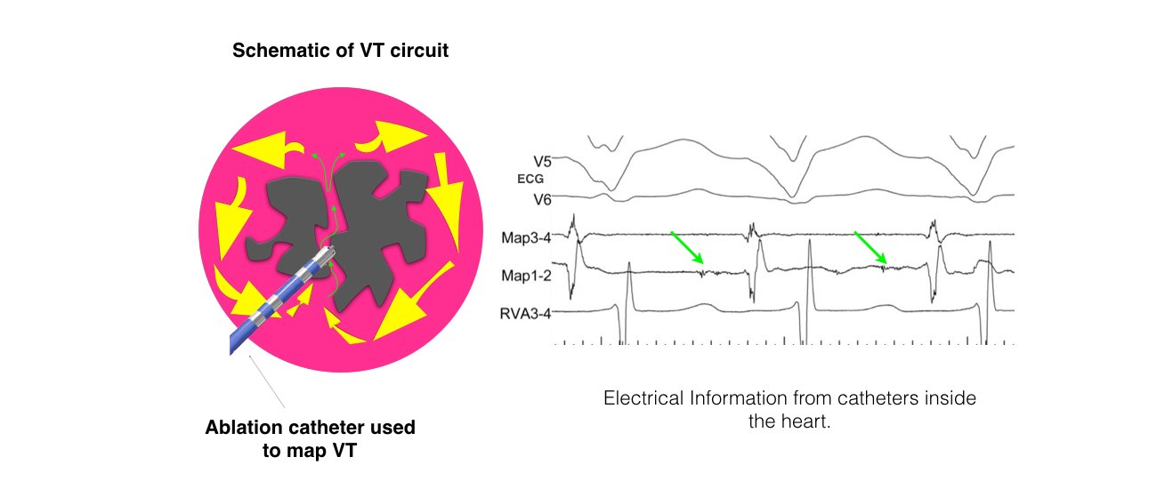
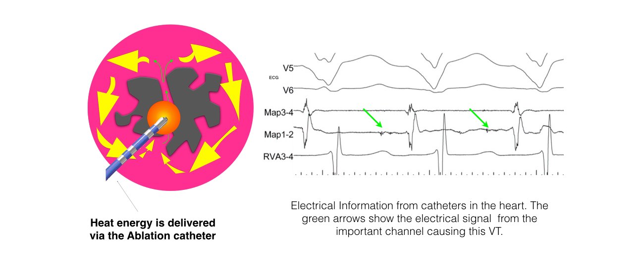
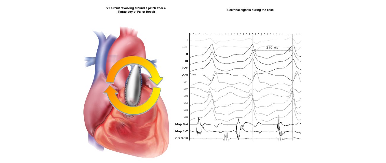
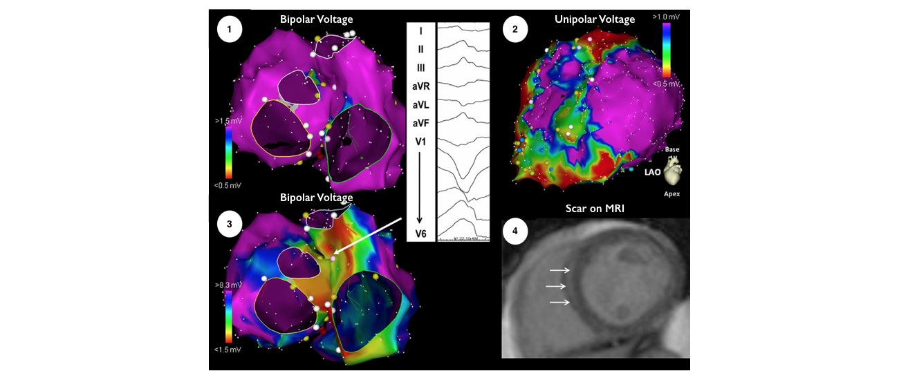
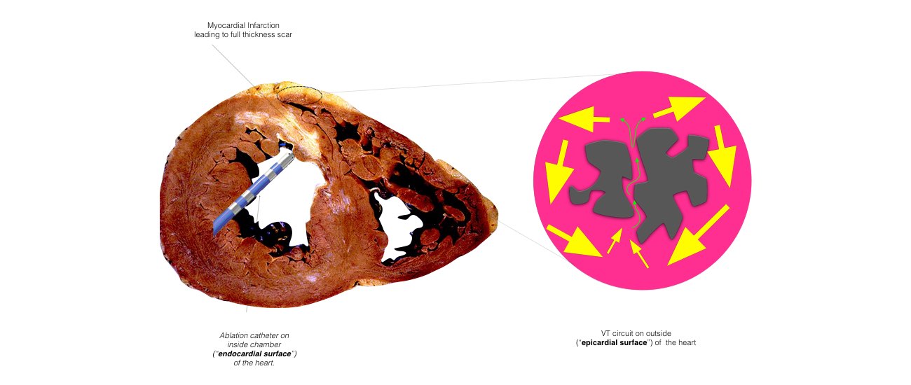
The catheters are positioned in your heart using X-Ray guidance. Once the catheters are in place you may feel your heart being stimulated and usually your abnormal heart rhythm will be induced. We will use a three-dimensional computer mapping system to guide the ablation procedure. This will help us move the catheters in your heart without the need for X-rays and also help us create an electrical map of the VT circuits. When the VT circuits have been identified and the abnormal tissue localized, the radiofrequency ablation will be applied to this spot. This may cause a transient warm discomfort in the chest. Radiofrequency ablation procedures are lengthy and the average duration is approximately 3 to 4 hours.

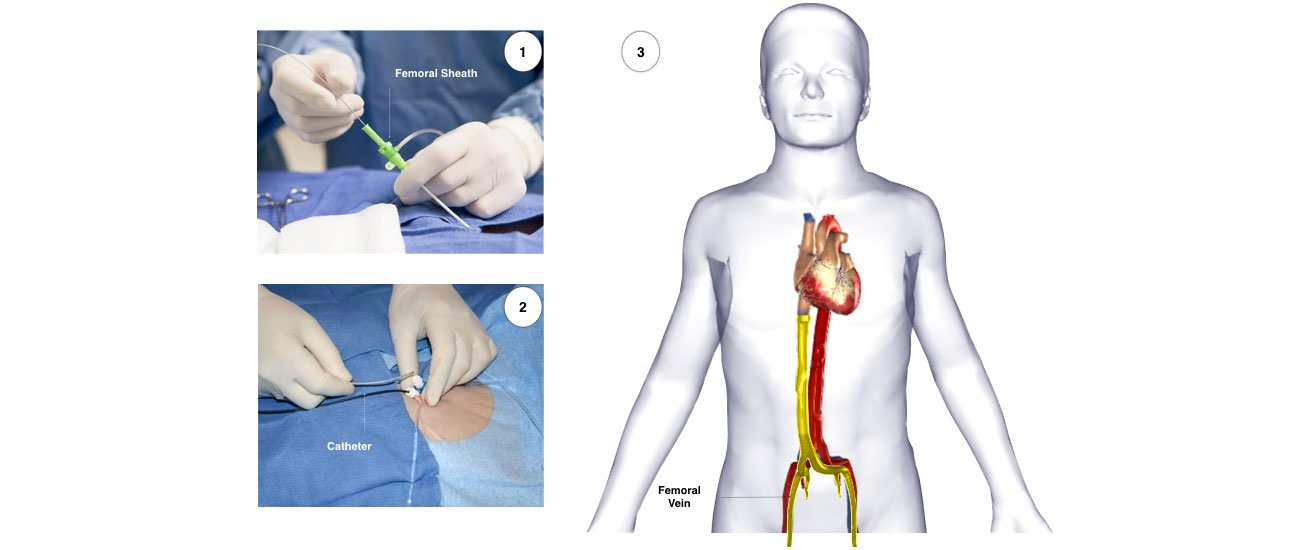
The catheters are inserted through intravenous ports, or sheaths, placed in the veins in the groin and sometimes through a vein on the side of the neck. To access the left ventricle, a needle may be used to create a small puncture in the wall between the right and left sides of the heart under ultrasound guidance (called transseptal catheterization). Alternatively, a catheter can be inserted into the heart through an artery in the groin (similar to heart catheterization procedures). The ablation catheter is moved around the ventricle, and a virtual 3-dimensional image of the heart is created with a computer mapping system that acts like a navigation system . The location of the catheter is determined by use of fluoroscopy (x-ray) and this mapping system. Typically, the procedure lasts from 3 to 6 hours.

  • The collage image shows the Radiofrequency Ablation Procedure.
  • A collage image of the reports also shows the scar on the MRI.

What is Epicardial VT ablation?

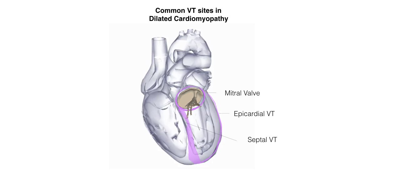
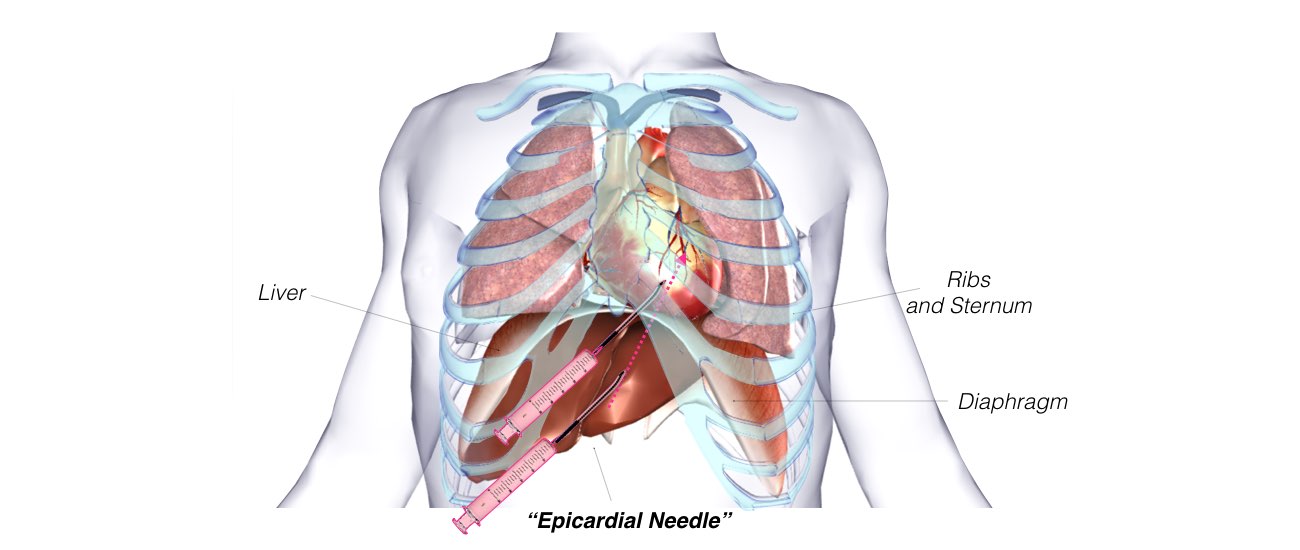
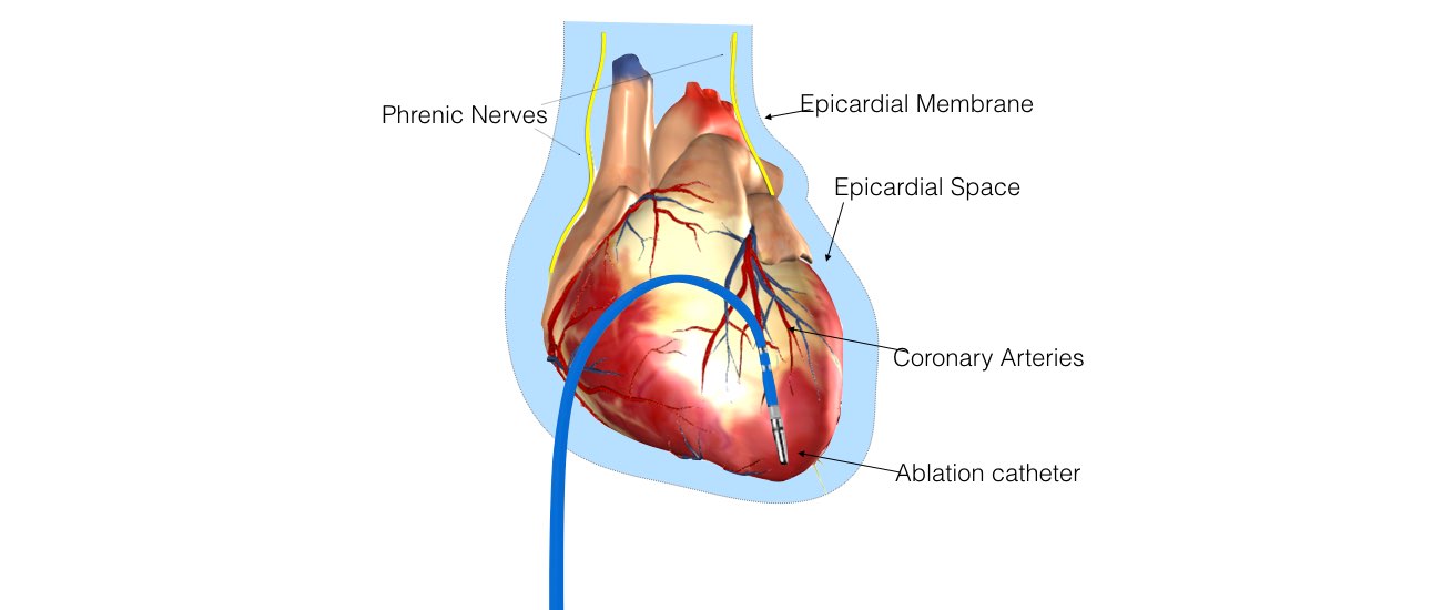
In some instances, the physician determines that the VT may originate from a circuit on the outer surface of the heart, or epicardium. If this is the case, a puncture into the sac, or pericardium, around the heart is performed just beneath the breastbone. This enables the ablation catheter to be inserted and maneuvered within the pericardium to determine whether the VT originates there. If a patient has a previous history of open heart surgery, a small surgical incision may be necessary to access the pericardium because of the presence of scar tissue, which can make the pericardium stick to the heart. These procedures to access the epicardium are usually performed at highly experienced centers.

What happens after the VT ablation procedure?

Afterward, the catheters are removed, but the sheaths are left in until the blood thinner wears off. Typically, this requires the patient to lie still for several hours to prevent bleeding from the puncture sites. Slight discomfort and bruising in the groin area can occur, and some patients experience self-limited mild chest pain resulting from inflammation caused by the ablation lesions. When the procedure is successful, antiarrhythmic medications may be stopped at the discretion of the physician.

  • The image shows the ablation catheter on the inside chamber of the heart and the VT Circuit on the outside of the heart.
  • The image shows the heart inside the rib cage and the epicardial needle used for Epicardial VT ablation.
  • The image shows the heart inside the rib cage and the ablation catheter in the VT ablation procedure.

RISKS/BENEFITS

What Are the Risks From the Procedure?

The risk of any major complication for VT ablation in patients with structural heart disease (Ischemic and Non-ischaemic Cardiomyopathy) is approximately 5%.

Major risks of VT ablation in structural heart disease include but are not limited to:

  • The peri-procedural risk of death during a VT 0.4 – 1.0%.
  • The risk of stroke or transient ischemic attack is 1-2% .
  • Vascular Complications < 1%.

The risks of epicardial access and epicardial ablation include but are not limited to:

  • Intra-abdominal or thoracic bleeding that can require surgery
  • Perforation and bleeding of the right ventricle.
  • Damage to the coronary arteries.
  • Damage of the phrenic nerve causing paralysis to one side of the diaphragm

How successful is a VT ablation?

The success of VT ablation varies, depending on the patient’s specific heart condition that caused VT. The procedure is most effective in patients with otherwise normal hearts, in whom the success rate exceeds 90%.

In patients with structural heart disease resulting from scar or cardiomyopathy, success rates upto 75% at 6 to 12 months. In cases when a patient experiences a recurrence, 2 of 3 patients will still have less VT than before the initial ablation.

-Senior Consultant Cardiologist & Electrophysiologist

-Chief, Cardiac Pacing and Arrhythmia Services

-Department of Cardiac Pacing and Electrophysiology

-Apollo Hospitals, Greams Road, Chennai.

© Arrhythmia Awareness Academy 2024 All Rights Reserved.
Designed by Digital SEO
chevron-down