Pediatric Pacemaker Implantation

BACKGROUND

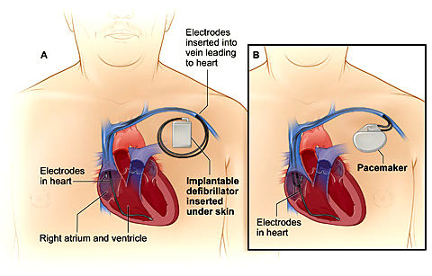
The Implantation of cardiac pacing devices in children and young adults can be challenging and different from the adult population due to their smaller size, their longer life expectancy, and anatomical variations associated with congenital heart defects.

Knowledge of indications, pacing leads and devices, anatomical variations, and the technical skills are important for those who implant and care for children with pacemakers.

Pacing in children is mainly performed in the setting of congenital or post-surgical complete heart block and less frequently in some surgical patients with sinus node dysfunction.

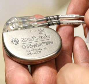
The indications, anatomical variations, and technical skills required for pacing children are different compared to those for adults. Also, current pacemakers are smaller sized, have longer battery life, multiple programming options, and therapeutic capabilities, and therefore provide greater options for pacing in children.

PROCEDURE INFORMATION

Implantation of Pacing Systems in Children

Pacing leads can be implanted via the transvenous (endocardial) or surgical (epicardial) route. The choice of route is dependent upon the size of the patient, anatomy, and surgical procedures performed that can affect the access to certain cardiac structures.

Epicardial Implantation

We recommend epicardial pacing in patients less than 10 kg, patients with intracardiac shunt lesions, patients with limited access to the atrium or the ventricle (e.g. patients with single ventricular physiology post Fontan palliation), and patients with prosthetic tricuspid valves.

Epicardial lead implantation requires sternotomy or thoracotomy or subxiphoid approach, and is associated with higher chronic stimulation threshold, higher lead failures and fractures, and early depletion of battery life. However, it preserves the venous access for future use.

Dual chamber epicardial pacemakers can easily be implanted in children over 3 kg.

Endocardial Implantation

Transvenous route of lead implantation is preferred in most patients, except for those situations referred above. Endocardial lead placement offers the advantages of avoidance of thoracotomy, lower pacing thresholds, and a lower incidence of lead fractures. However, its disadvantages include a greater risk of lead dislodgment and venous occlusion.

FOLLOW-UP

The patient and the device are assessed prior to discharge, in 1-2 weeks for an incision check, at 2-3 months to assess chronic pacing thresholds and cardiac function (because of the risk of pacing-induced cardiac dysfunction), and then every 6 months in infants and children.

-Senior Consultant Cardiologist & Electrophysiologist

-Chief, Cardiac Pacing and Arrhythmia Services

-Department of Cardiac Pacing and Electrophysiology

-Apollo Hospitals, Greams Road, Chennai.

© Arrhythmia Awareness Academy 2024 All Rights Reserved.
Designed by Digital SEO
chevron-down