Left Atrial Appendage (LAA) Closure

Background

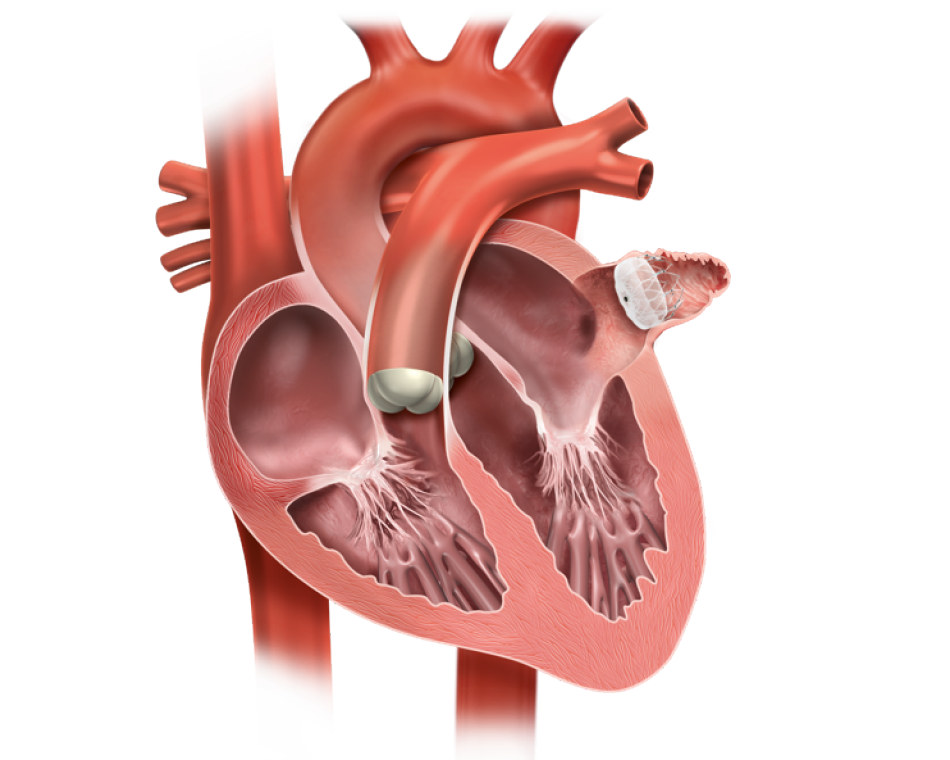
Left atrial appendage closure is a procedure that closes off the opening of your left atrial appendage. Your left atrial appendage is a small pouch, shaped like a windsock, found in the top left of your heart (the left atrium). Like your appendix, your left atrial appendage doesn’t really have a clear role to play in your body.

Why do I need a left atrial appendage closure?

If you have a disease known as atrial fibrillation – which occurs when your heart’s electrical system is disturbed and your heartbeat becomes irregular.

As your heart contracts with each heartbeat, blood is squeezed out of the left atrium and into the left ventricle (bottom left of your heart). If you have atrial fibrillation, blood can’t be squeezed out of your left atrium effectively, and can therefore collect in your left atrial appendage and increase your chances of a stroke.

Blood thinning medications are a common treatment for atrial fibrillation but they aren’t suitable for everyone. If you can’t take medication, left atrial appendage closure is an alternative option to prevent stroke.

  • Close-up image of a special catheter in the heart.
  • A bisection image of the heart showing the catheter.

Pre/Post Procedure

You will prepare for your left atrial appendage closure by:

Transoesophageal Echocardiogram (TOE) – You have to take TOE pictures of your heart to make sure there are no clots before your procedure commences.

What happens during a left atrial appendage closure?

Left atrial appendage can be closed through your skin without requiring surgery. The procedure takes place in a cath lab, and you’ll will be connected to heart monitors throughout the procedure. During this percutaneous procedure:

  • A cannula will be put in your arm to give you medication
  • A local anaesthetic to numb the area – or a general anaesthetic may be given – so you don’t feel any discomfort
  • A special catheter is gently inserted in your groin and guided slowly up to your heart across the wall that separates the right and left side of your heart
  • Using X-rays, X-ray dye and TOE, a device is then positioned and expanded at the opening of the left atrial appendage (this acts like a seal or “plug” and keeps clots from being released)

The catheter is removed and the device stays in. Over time a thin layer of tissue will grow over the surface of the device

-Senior Consultant Cardiologist & Electrophysiologist

-Chief, Cardiac Pacing and Arrhythmia Services

-Department of Cardiac Pacing and Electrophysiology

-Apollo Hospitals, Greams Road, Chennai.

© Arrhythmia Awareness Academy 2024 All Rights Reserved.
Designed by Digital SEO
chevron-down