BACKGROUND

An EP study is a low-risk procedure that has been performed in major medical centres for many years. The EP study makes it possible to study your abnormal heart rhythm under controlled conditions and diagnose your particular problem. (In this procedure, one or more thin, flexible wires, called catheters, are inserted into a blood vessel usually the groin and guided into the heart). Each catheter has two or more electrodes to measure the heart’s electrical signals as they travel from one chamber to another. An EP study is usually done under local anaesthesia.

An EP Study offers more detailed information about the heart’s electrical activity than many other noninvasive tests because electrodes are placed directly on heart tissue. EP studies are done to diagnose your cardiac rhythm abnormality, especially in a patient with palpitation or unexplained loss of consciousness, to help determine the best treatment. EP study allows the Electrophysiologist to determine the specific location of an arrhythmia and, often, to correct it during the same procedure.
PROCEDURE INFORMATION

Who requires EPS?

Patients are referred for EPS for many reasons.

Some of the more common reasons are:

  • Rapid or irregular heartbeat (often associated with shortness of breath, chest pain or dizziness). Common terms used are palpitations, racing heart or missed beats.
  • Blackout or dizziness.
  • Abnormal findings on an ECG.

Normal heart rhythm.

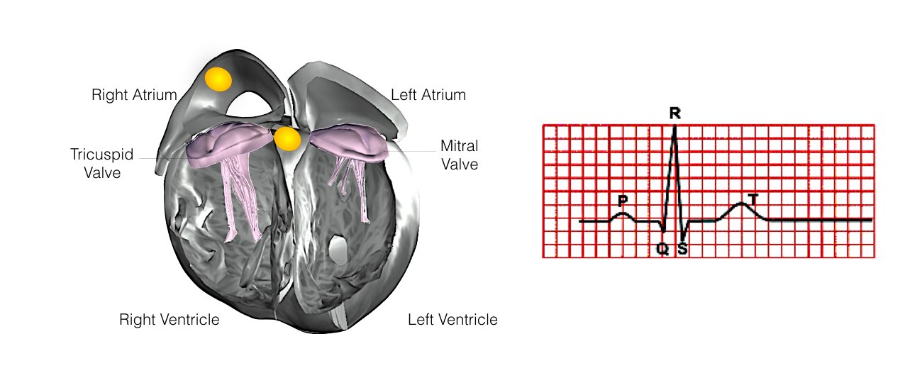
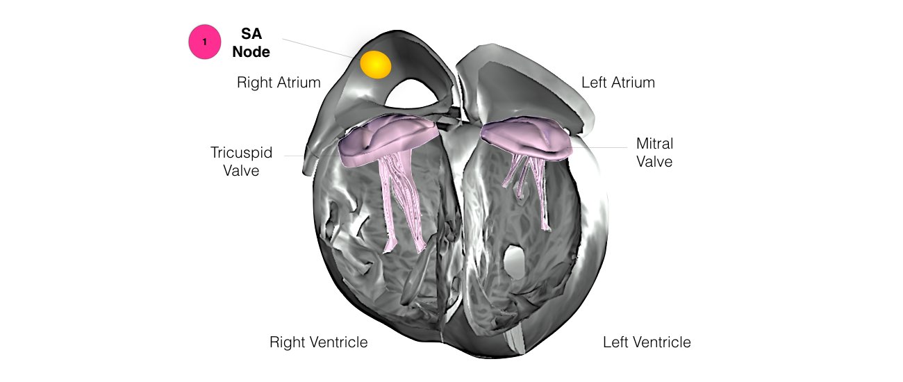
In order for the heart to do its work (pumping blood throughout the body), it needs a sort of spark plug or electrical impulse to generate a heartbeat. Normally, this electrical impulse begins in the upper right chamber of the heart (in the right atrium) in a place called the sinoatrial (SA) node. The SA node is the natural pacemaker of the heart. The SA node gives off electrical impulses to generate a heartbeat in the range of 60 to 100 times per minute.

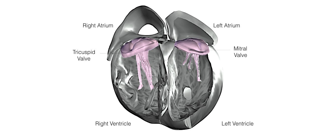
  • The internal structure of a heart.
  • The internal structure of a heart shows the SA node.
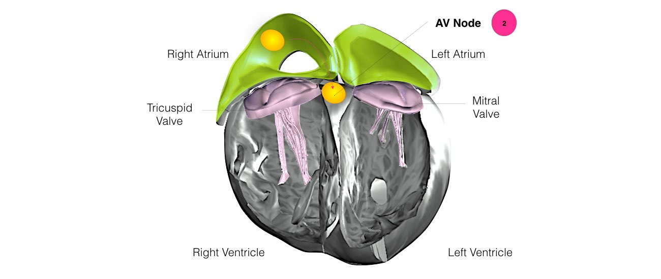
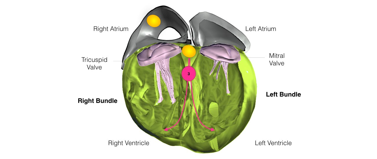
  • The internal structure of a heart shows the AV node.
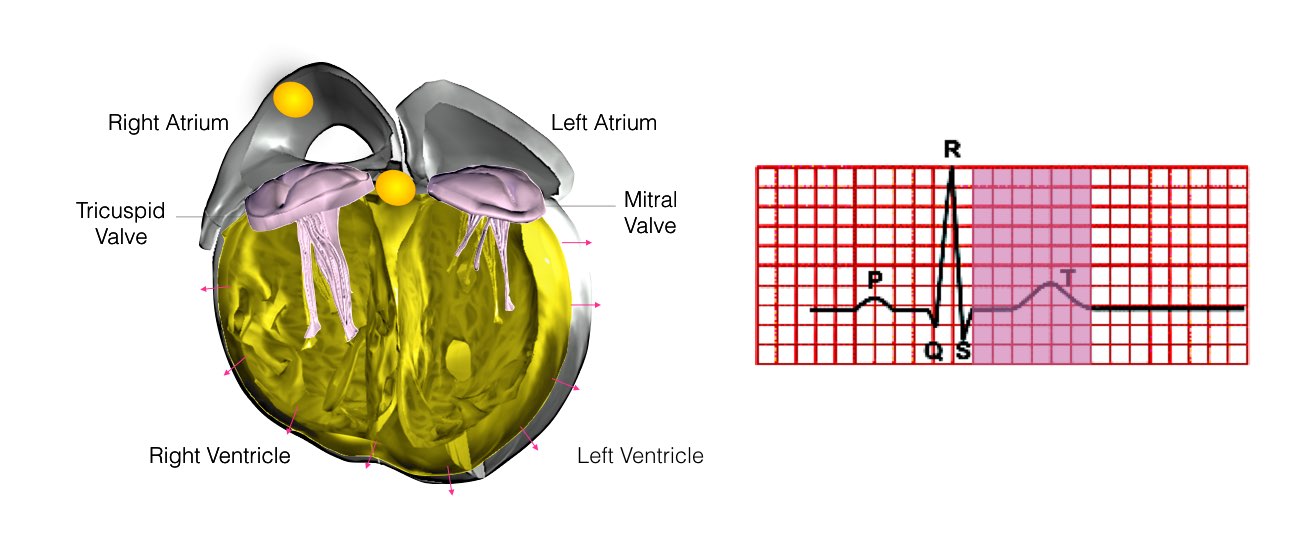
  • Abnormal heart rhythm at the expansion of the lower chambers with ECG reading.
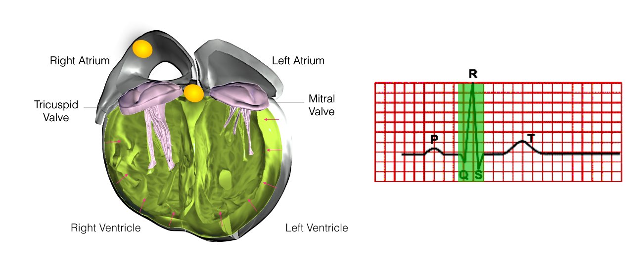
  • The image shows the internal heart structure at the contraction of the lower chambers with ECG reading.
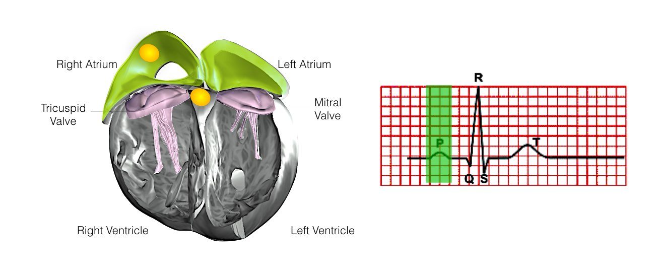
  • Abnormal heart rhythm at the upper chambers with ECG reading.
  • The internal structure of a heart shows the SA and AV nodes with ECG reading.
  • The image shows the electrical impulses from the AV node.

How do abnormal heart rhythms occur?

In some hearts, an abnormal heart rhythm develops when an electrical impulse either starts from a different location, other than the SA node, or follows a route (or pathway) that is not normally present.

Your doctor will explain the exact nature of your abnormal heart rhythm and the following diagrams should help you understand the mechanism.

Types of abnormal heart rhythms include:

1. AV Nodal Re-entry Tachycardia (AVNRT)

This is the most common form of SVT. An abnormal short circuit (circular conduction) occurs near the AV node. Instead of a single AV node between the top and bottom chambers, there is a second connection that is abnormal. This extra connection has been present since birth. As a result of having 2 connections, a short-circuit can occur.

2. Re-entrant Tachycardia and Wolff Parkinson White Syndrome

Abnormal circular conduction utilising the AV node and an “accessory pathway” connecting the atria and ventricles. This extra connection has been present since birth. As a result of having 2 connections a short-circuit can occur. This condition is sometimes termed the Wolff-Parkinson-White Syndrome or WPW.

3. Atrial Tachycardia

This is the least common form of SVT. There is an extra abnormal origin of the electrical impulse from a small area in the atria other than the SA node. It is not known when or why such an extra focus develops.

4. Atrial Flutter

In atrial flutter, abnormal electrical signals circulate in the heart’s upper chambers, or atria, which causes the rhythm of the lower chambers to be fast. Because contractions are not timed as in the normal heartbeat, the heart does not pump blood as effectively to the rest of the body. Irregular heartbeats can be seen on an electrocardiogram, or ECG

5. Atrial Fibrillation

In atrial fibrillation, disorganized electrical signals originate in the heart’s upper chambers, or atria, causing the rhythm to be irregular. These signals come from small areas in the heart, represented here by stars. Irregular heartbeats can be seen in an Electrocardiogram or “ECG”. Because contractions are not coordinated as in the normal heartbeat, the heart does not pump blood effectively to the rest of the body.

6. Ventricular Tachycardia

Ventricular Tachycardia is a heart arrhythmia, or irregular heartbeat. In ventricular tachycardia, abnormal electrical pulses in the lower chambers, or ventricles, disrupt the normal firing of the SA node, causing the heart to beat rapidly. These signals can be seen in an Electrocardiogram or ECG. A rapid heartbeat does not give the heart enough time to refill with blood before pumping, which causes diminished blood flow to the rest of the body. This may lead to symptoms of dizziness, lightheadedness, and unconsciousness.

PRE/POST PROCEDURE

What happens prior to your procedure?

  • You will receive an admission letter outlining the date of your procedure.
  • You will be asked to discontinue some of the anti-arrhythmic medications 2-3 days prior to the procedure.
  • If you are taking anticoagulation (blood thinning) medication e.g. Warfarin, then you will need to stop this for 5-7 days prior to your procedure.
  • You will be required to fast for at least six hours before the EP study.

What happens during an EP study?

  • You will be transferred to the Electrophysiology Laboratory (EP lab) from your ward. Usually before leaving your ward, your groin will be shaved.
  • The EP lab has a patient table, X-Ray tube, ECG monitors and various equipment. The staff in the lab will all be dressed in hospital theatre clothes and during the procedure will be wearing hats and masks.
  • Many ECG monitoring electrodes will be attached to your chest area and patches to your chest and back. These patches may momentarily feel cool on your skin.
  • A nurse or doctor will insert an intravenous line usually into the back of your hand. This is needed as a reliable way to give you medications during the study without further injections. You will also be given further sedation if and as required. You will also have a blood pressure cuff attached to your arm, which will automatically inflate at various times throughout the procedure.
  • The oxygen level of your blood will also be measured during the EP study and a small plastic device will be fitted on your finger for this purpose. Your groin area and possibly your neck or arm will be washed with an antiseptic cleansing liquid and you will be covered with sterile sheets, leaving these areas exposed.
  • The doctor will inject local anaesthetic into the area where the catheters are to be placed. After that, you may feel pressure as the doctor inserts the catheters, but let the staff know if there is any discomfort so some more local can be given. Once the catheters are in place, you may feel your heart being paced and possibly your abnormal heart rhythm will be induced.
What to expect after your procedure?
  • After procedure you will be transferred back to your ward where you will have to lie flat for 4-6 hours depending on which blood vessels have been punctured. During this time, it is important to keep your legs straight and your head relaxed on the pillow.
  • Most patients stay in hospital overnight and their heart rhythm may be monitored during this time.
  • The majority of patients have 2-3 days away from work.

RISKS/BENEFITS

What risks are involved in an EP study?

The EP study is a very low-risk procedure. The worldwide complication rate for EP study is less than 0.5%.

-Senior Consultant Cardiologist & Electrophysiologist

-Chief, Cardiac Pacing and Arrhythmia Services

-Department of Cardiac Pacing and Electrophysiology

-Apollo Hospitals, Greams Road, Chennai.

© Arrhythmia Awareness Academy 2024 All Rights Reserved.
Designed by Digital SEO
chevron-down