BACKGROUND

The heart is a pump responsible for maintaining blood supply to the body. It has four chambers. The two upper chambers (the right atrium and left atrium) are the chambers that receive blood as it returns from the body via the veins. The lower chambers (the right and left ventricle) are the chambers responsible for pumping the blood out to the body via the arteries. Like any pump, the heart has an electrical system that controls how it functions.

Normal heart rhythm.

In order for the heart to do its work (pumping blood throughout the body), it needs a sort of spark plug or electrical impulse to generate a heartbeat. Normally this electrical impulse begins in the upper right chamber of the heart (in the right atrium) in a place called the sino-atrial (SA) node. The SA node is the natural pacemaker of the heart. The SA node gives off electrical impulses to generate a heartbeat in the range of 60 to 100 times per minute. If you are exercising, doing strenuous work or you are under a lot of stress, your heart rate may be faster. When you rest or sleep your heart rate will slow down. If you take certain medications, your heart rate may be slower. All of this is appropriate.
From the SA node, the electrical impulse is relayed along the heart’s conduction system. It spreads throughout both the right and left atria causing them to contract evenly.
When the impulse spreads over the right atrium it reaches the atrio-ventricular (AV) node. This is a very important structure in the heart because it is the only electrical connection between the top chambers and the bottom chambers. It is therefore the only way in which an electrical impulse can reach the pumping chambers (the ventricles). The impulse spreads through the AV node and down into the lower chambers or ventricles of the heart. This causes them to contract and pump blood to the lungs and body.

Abnormal Conduction Develops in Cardiomyopathy Patients.

Some patients with cardiomyopathy develop an abnormality in their conduction system. The most common abnormality is a delay in the normal electrical conduction through one of the conducting wires in the ventricle – the left bundle branch. This is known as the left bundle branch block (LBBB) and can be diagnosed on the ECG as a widening of the QRS (>120ms) complex. This is seen in approximately 40% of patients with cardiomyopathy. Because of the delay in conduction down the left bundle branch, the right ventricle is activated a fraction of a second earlier than the left ventricle. Additionally the LBBB causes the lateral wall of the left ventricle to be activated much later than normal. This dyssynchrony leads causes an overall decrease in the pump function of an already weakened heart in heart failure patients. Patients with LBBB on ECG and heart failure symptoms may benefit from Cardiac Resynchronisation Therapy (CRT), this is also called a Bi-Ventricular Pacemaker.
  • The internal structure of a heart shows the AV node the delay in conduction down the left bundle branch.
  • The internal structure of a heart shows the Right Ventricle, left ventricle, right atrium, and right atrium.
  • The internal structure of a heart shows the SA node.
  • The image shows impulse spreads through the AV node and into the upper chambers.
  • The image shows impulse spreads through the AV node and down into the lower chambers with ECG reading.
  • The image shows the AV node and the contraction of lower chambers with ECG reading.
  • The image shows impulse spreads through the AV node and down into the upper chambers with ECG reading.
  • The image shows the internal structure of the heart with ECG reading.
  • The image shows an abnormality in the electrical conduction system.
What is Cardiac Resynchronization Therapy (CRT)?
The aim of CRT is to restore the normal coordinated pumping action of the ventricles by overcoming the delay in electrical conduction delay caused by the left bundle branch block. This is achieved by implantation of a specialized pacemaker called a CRT device or Bi-Ventricular pacemaker. In addition to the standard wires (leads) that are implanted in the right atrium and right ventricle present in a normal dual chamber pacemaker, a third wire (lead) is implanted to stimulate the lateral wall of the left ventricle. We can pace the lateral wall of the left ventricle via the coronary sinus which is a vein which wraps around the back of the heart, around the left ventricle. This vein can be accessed via the right atrium.
Once the LV lead is positioned, the CRT device will be programmed to pace the right ventricle and left ventricle simultaneously. This leads to improved coordination of the heart’s pump function and can be seen as a narrowing of the QRS in the ECG. Studies have shown CRT leads to improved exercise tolerance and improved quality of life.
Who is a candidate for CRT?

Patients who will benefit from a CRT device include those with:

  • Heart failure and moderate to severe heart failure symptoms (decreased exercise tolerance and shortness of breath) despite optimal heart failure medications.
  • Cardiomyopathy (weakened and enlarged heart muscle).
  • Significant left bundle branch block.

Some patients with cardiomyopathy who are candidates for CRT are also at high risk of sudden cardiac death from dangerous heart rhythms originating from the ventricles (Ventricular Tachycardia and Ventricular Fibrillation). For these patients a special CRT device is available that can stop these dangerous heart rhythms delivering a shock known as a defibrillator. These devices incorporate a standard Implantable Cardioverter Defibrillator (ICD) with a CRT pacemaker and are called CRT-D devices. The choice of a CRT versus and CRT-D is dependent on many clinical factors and will be discussed with you.

Procedure Information

The device is inserted just under the collar-bone on the front of the chest, usually on the left side. The area is prepared with a special sterile solution that may feel cold. You will be covered by a large sterile sheet that will partly cover your face. You will be able to look out from under the sheet to the side and a nurse will be present at all times. You will be given oxygen to breathe by a small tube that is positioned under your nostrils or by a mask that covers your face.

At the start of the procedure, the doctor will inject local anaesthetic into the area under the collar-bone where the CRT device is to be inserted. This will sting momentarily but the area will then be numb. During the procedure you may feel some firm pushing in the shoulder area but this should not be painful. If you experience pain or discomfort you should tell the nurse or doctor.

Three leads (special wires which connect the CRT device with the heart) are inserted into the heart via the vein under the collarbone. These are manipulated into the heart under X-Ray control. Initially the standard leads are inserted first, one into the right atrium and one into the right ventricle. These leads attach to the heart wall either with small hooks or with a small screw. The coronary sinus is accessed using a small thin tube (called a sheath) and the left ventricular lead is passed via the sheath into a lateral branch of the coronary sinus. All three leads will then be connected to the CRT device.

Sometimes it is not possible to pass the third wire into the coronary sinus due to its shape. A possible option in these situations is referral to a cardiothoracic surgeon to implant the lead via a limited open surgical approach at a second procedure. This will be discussed with you if the LV lead cannot be implanted successfully.

  • The image shows a heart with an implanted CRT pulse generator.
  • The lateral and posterior view of the heart after implanting the CRT pulse generator.
  • Picture of a heart with the right atrial lead and left atrial lead.
  • The image illustrated injecting a local anesthetic into the area under the collarbone where the CRT device is to be inserted.
  • The image illustrates the Cardiac Resynchronization procedure.

PRE/POST PROCEDURE

What happens prior to your procedure?

If you are taking anti-coagulation (blood thinning) medication e.g. warfarin then you will need to stop this for approximately 5 days prior to your procedure. Your doctor may arrange for you to have daily heparin injections after you stop the warfarin.

Patients will be required to attend the pre-admission clinic on the day prior to the procedure.

At the pre-admission clinic you will see a doctor who will record your medical history. You will also require an ECG and blood test. The doctor will also confirm the time you should be at the hospital for admission the following day.

You will be required to fast for at least six hours before each of the procedures. If your procedure is in the afternoon you may have a light breakfast. If your procedure is in the morning, DO NOT EAT OR DRINK AFTER MIDNIGHT, except for sips of water to help you swallow your pills.

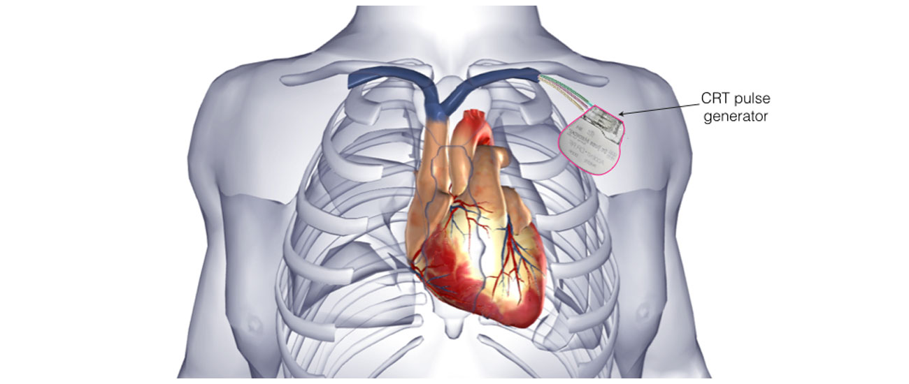
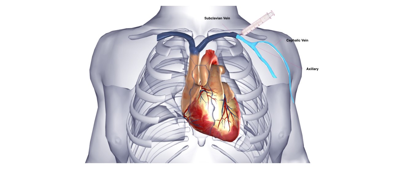
Insertion of a CRT device is now a very common procedure. This is performed either under local anaesthetic with sedative medication to make you feel comfortable or under general anaesthetic. Your doctor will discuss this with you. The procedure takes approximately 2 hours and is performed in the cardiac catheter laboratory.

This is a special room that has a patient table, X-Ray tube, ECG monitors and other equipment. The staff in the lab will all be dressed in hospital theatre clothes and during the procedure will be wearing hats and masks.

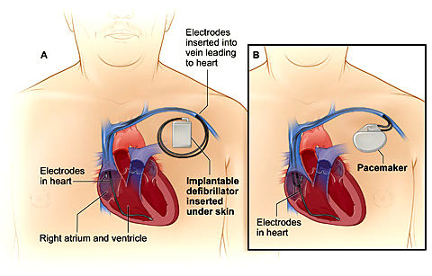
Many ECG monitoring electrodes will be attached to your chest area. A nurse or doctor will insert an intravenous line usually into the back of your hand. This is needed as a reliable way to give you medications during the study without further injections. You will also have a blood-pressure cuff attached to your arm that will automatically inflate at various times throughout the procedure.

What happens after CRT Implantation?

After the procedure you will have some bruising and discomfort in the area of the CRT device that may persist for several weeks. This bruising can create a bluish discoloration over the upper chest and arm. This is normal.

You should avoid strenuous activities with your arm or from lifting the arm above your head for a period of 4 weeks. You should refrain from driving for 2 weeks. If you have already had a heart rhythm disturbance you may be disqualified from driving for 6 months. Your doctor will discuss this with you. You should not go swimming, play golf, or bowling for 4 weeks.

A sterile dressing is left over the pacemaker for 8 days. You can carefully remove this dressing yourself on the 8th day after the procedure. At this stage the wound is sufficiently healed to allow you to shower with the dressing removed.

You will be allowed to go home 1 or 2 days after the procedure.

You will be given an appointment to see the doctor 1 month after the implant.

What should I do if I have concerns after the implantation?

Usually the discomfort and swelling from the wound settles gradually over several weeks. If the wound becomes increasingly tender, reddened and swollen or you have any other concerns, you should contact your cardiologist.

RISKS/BENEFITS
What are the benefits of CRT?
The response to CRT can vary between patients. Published studies involving several thousand patients worldwide have demonstrated improvements in exercise tolerance, heart failure symptoms and quality of life. It often takes weeks to months for these improvements to be noticed. Unfortunately a small number of heart failure patients do not benefit from the CRT therapy.
What are the Risks of CRT Insertion?

CRT implantation is a very common and low risk procedure and should a complication arise, it will be dealt with at once.

Although most people undergoing CRT implantation do not experience any complications, you should be aware of the following risks:

  • Haematoma (large bruise) – this may occur at the pacemaker insertion site. This may be uncomfortable and can take several weeks to settle.
  • Pneumothorax –During the procedure it is necessary to insert the pacemaker leads into your heart via a small vein under the collar-bone. This vein runs very close to the lung and there is a small chance (1 in 100) that a small hole could be inadvertently made in the lung (Pneumothorax). Should this occur, it would usually heal by itself. However, occasionally a small tube may need to be inserted to drain out the air. This can be uncomfortable and means spending several extra days in hospital.
  • Lead Dislodgement. – Although a great deal of care is taken in placing the pacemaker leads inside your heart, occasionally one of them moves and will need to be repositioned. The risk of lead dislodgement is approximately (1 in 40). This usually occurs in the first 24 hours after the procedure and is detected by testing the pacemaker.
  • Infection – There is a very small chance (1 in 100) that the pacemaker will develop an infection. Should this occur, it is usually necessary to remove the pacemaker in order to clear the infection.
  • Cardiac perforation – Very rarely (1 in 1000), one of the leads can make a small hole in the heart causing blood to accumulate around the heart. If this occurs the problem will be dealt with immediately. This is a very rare occurrence.
  • Subclavian Vein Thrombosis- A blood clot may form in the subclavian vein. This is the vein that the leads go through to get to the heart

-Senior Consultant Cardiologist & Electrophysiologist

-Chief, Cardiac Pacing and Arrhythmia Services

-Department of Cardiac Pacing and Electrophysiology

-Apollo Hospitals, Greams Road, Chennai.

© Arrhythmia Awareness Academy 2024 All Rights Reserved.
Designed by Digital SEO
chevron-down